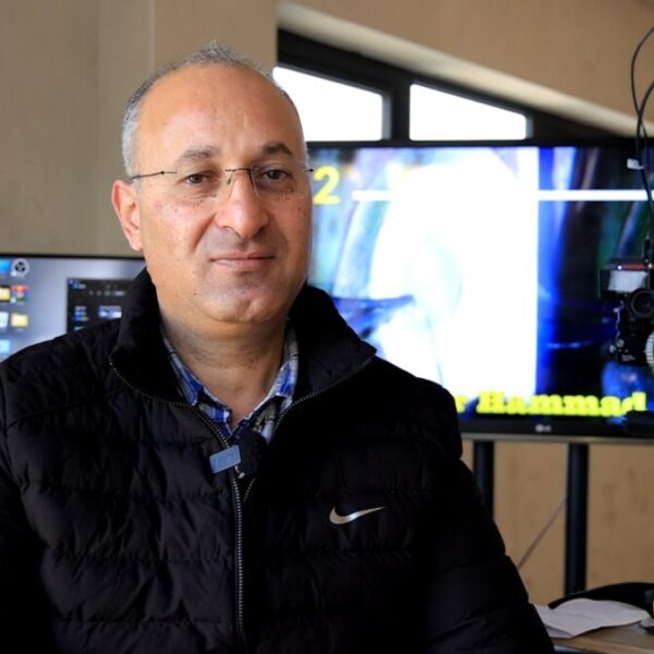
As a Palestinian proverb says, "necessity is the mother of invention." A principle that Dr. Omar Hamad, a Palestinian ophthalmologist, has embraced. Specializing in eye care, he developed a 3D microscope by adapting existing standard equipment, using his own resources and skills. This initiative aims to improve surgical precision and working conditions for doctors, while making 3D technology accessible in a context marked by limited resources.
AI Index: Mediterranean Knowledge Library
An ophthalmologist develops a 3D microscope from a classic device
22-med – January 2026
• In Palestine, a doctor adapts a classic ophthalmic microscope to transform it into a high-performance, low-cost 3D device.
• A local medical innovation that improves surgical precision and opens up modernization prospects for health services.
#palestine #health #innovation #medicine #ophthalmology #technology #research #autonomy #mediterranean
This innovation is concretely changing surgical practice in ophthalmology by making accessible a technology that previously depended on very expensive equipment.
Dr. Omar Hamad's motivation stems from an international conference where a foreign professor refused to answer his technical questions about a 3D microscope and suggested he buy the device instead of trying to understand it. "I felt that Arabs were perceived as incapable of innovating. It was at that moment that I decided to develop my own system. I spent a year attending scientific conferences and specialized publications to understand how commercial 3D systems work," he states.
By transforming a standard microscope, manufactured in Germany and widely available on the Palestinian market, into a functional 3D device, Dr. Omar Hamad achieved results comparable to those of commercial models valued at nearly 275,000 euros ($300,000), for a modification cost of about 18,500 euros ($20,000).
The system enhances depth perception, the surgeon's comfort, and the precision of movements, while allowing the medical team to monitor procedures in real-time. Initial trials conducted on animal eyes show an increase in operational ease and the likelihood of surgical success. They thus pave the way for large-scale modernization of existing equipment without relying on imported and financially out-of-reach solutions.
From Existing Equipment to 3D Imaging
The development of the microscope took place in two distinct phases. The first involved integrating additional equipment and testing the device, while the second focused on conducting examinations and surgical procedures on animals. "The first phase checks technical feasibility, while the second tests surgical maneuvers in conditions close to reality," the ophthalmologist specifies.
The modifications required the purchase and installation of cameras, screens, chips, and 3D glasses. "Once the system was installed, depth visualization became much clearer," he explains, referring to both increased physical and psychological comfort during work.
The 3D images were then merged via a high-speed computer system, linking two screens side by side to replicate the configuration of commercial 3D microscopes. The installation of 3D imaging software yielded results comparable to those of high-end devices.
The second phase, currently underway, relies on experimental surgeries conducted on animal eyes, particularly fish*. "The preliminary results are very positive," he states, indicating that the system allows procedures to be performed more easily and increases the likelihood of surgical success.
Overcoming Technical and Financial Constraints
However, the development of the microscope has not been without obstacles. The lack of sponsors for scientific research led him to personally finance all the equipment. The limited availability of specialized components forced him to spend long hours online negotiating with manufacturers, often reluctant to deal with individuals rather than companies.
Technical challenges, particularly latency in video transmission, were resolved pragmatically. "I used mobile phones to precisely measure delays and ensure high-precision 3D imaging," adds Dr. Omar Hamad.
Videos from the experimental procedures show clear and overlaid 3D images of the eye, confirming the effectiveness of the modified microscope. "Working with this system allows for better control without excessive physical effort," he emphasizes, while also enabling the medical team to monitor the intervention in real-time.
A Project Focused on Public Health Service
Installed in his eye care and surgery center, still under construction in Bethlehem, the modified microscope now occupies the main room. "I consider this initiative a major achievement, given the modest resources involved," he emphasizes. He hopes that this success, which he describes as Palestinian and Arab, will contribute to advancing scientific research and fostering innovative initiatives.
The goal is not to commercialize the device. "It is primarily about improving ophthalmology services in Palestine and the Arab world," he insists, highlighting the potential to convert existing standard microscopes into 3D systems.
Once all trials are completed, he plans to present his work to Palestinian medical authorities, the Ministry of Health, and the press. "I hope this solution can be adopted to modernize all microscopes used by the ministry to 3D capability," he concludes.
* Fish eyes are commonly used in this type of testing due to the optical simplicity of their lens, whose spherical shape facilitates observation and evaluation of depth visualization systems.
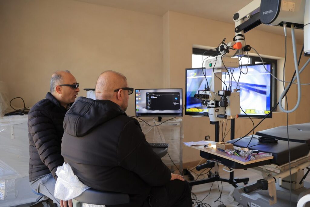
Cover photo: Dr. Omar Hamad © Ahmed Jubran